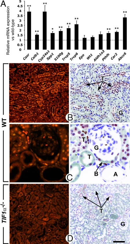
Fig. 3.
TIF1α is ubiquitously expressed in nephrons and represses gene expression in kidneys. (A) Total RNA extracted from kidneys of 3-month-old WT and TIF1α−/− mutants (n = 4 in each group) was analyzed by quantitative RT-PCR. Expression of the indicated genes was analyzed in triplicate together with HPRT, and results are represented as expression relative to HPRT for WT and mutant, with expression of each gene arbitrarily set equal to one for WT samples (indicated by the horizontal dotted line). *, P < 0.05; **, P < 0.01. (B–D) Histological sections through the kidney cortex immunostained with an antibody specific to TIF1α. Genotypes are as indicated. In D note the absence of stained nuclei in the TIF1α-deficient kidney. A, arteriole; B, Bowman's capsule of a glomerulus; G, kidney glomeruli; T, kidney tubules. (Scale bar: B and D, 80 μm; C, 20 μm.)

