
Figure 1. The Dmef2-5x[C/D]*-GAL4 driver causes expression of a GFP reporter in the somatic mesoderm and body wall muscles.
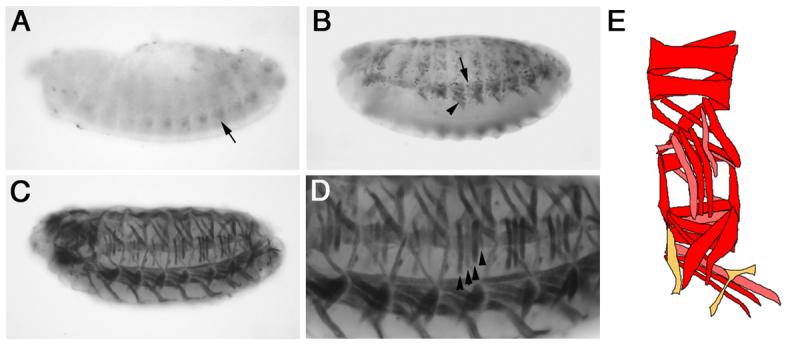
Dmef2-5x[C/D]*-GAL4 > UAS-GFP embryos were stained with an antibody against GFP. Lateral views of stage 14 (A), 15 (B) and 16 (C-D) embryos are shown. Panel D shows a close up of the embryo in panel C. (A) In contrast to the direct enhancer-reporter construct (Duan et al, 2001), GFP expression is first detected in a subset of cells in the ventral somatic mesoderm at during stage 14 (black arrow, A). (B) During stage 15, GFP expression is detected in growing myotubes (black arrowhead, B) and surrounding cells (black arrow, B). (C-D). GFP expression is detected in the final body wall muscles at stage 16. GFP is not detected in all muscles or at uniform levels. Black arrowheads highlight differing levels of GFP expression in LTs1-4. (E) Schematic showing quantification of GFP expression in individual muscles from Table 1. Red = 75-100%, pink = 50-74% and orange = <50%.