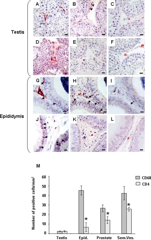
Figure 1. Localization and quantification of SIV/HIV target cells in the male genital tract.
Testis (A–F) and epididymis (G–L) immunolocalization of HLA-DR (A, G), CD68 (B, H), CD3 (C, I), CD4 (D, J), CCR5 (E, K) and CXCR4 (F, L) positive cells in uninfected macaques. Arrows show immunopositive cells in contact with the epithelium of the epididymis. Note the presence of testicular macrophages within the peritubular wall bordering the seminiferous tubules of the testis (Figure 1H, arrow heads).Scale bars = 20 µm. (M): Quantification of HIV potential target cells (CD68+ and CD4+ stained positive cells) in the male reproductive organs of uninfected macaques. Stars indicate statistical difference between the number of CD68+ cells and CD4+ cells within an organ (Wilcoxon signed rank test, p<0.05;). The number of CD68+ cells was significantly lower in the testis when compared with the other MGT organs (Wilcoxon signed rank test, p<0.05, not shown on the graph). The number of CD4+ cells in the testis was significantly lower than in the prostate and seminal vesicles (Wilcoxon signed rank test, p<0.05, not shown on the graph).

