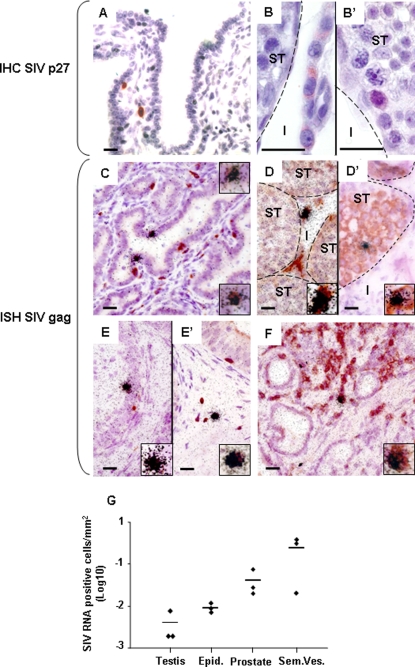
Figure 4. SIV localization within the male genital tract.
Detection of SIV positive cells in the seminal vesicles (A, C), testes (B–B', D–D'), epididymides (E, E') and prostate (F) of primary-infected macaques using immunohistochemistry for SIVp27 (A, B) and in situ hybridization (ISH) for SIV gag RNA (C–F). The phenotype of SIV positive cells was determined using ISH for SIV gag RNA (visualized as black silver grains) combined with immunostaining for cell markers (visualized as brown staining): combined ISH for viral RNA and immunostaining for CD3 revealed black silver grains clustered over brown cells in the seminal vesicles (C), prostate (F), and epididymis (E'), indicating infection of CD3+T lymphocytes. Co-labelling of SIV RNA+ cells with the myeloid cell marker CD68 was also observed, as shown here for the epididymis (E). In the testis, SIV RNA was detected within the interstitium in HLA-DR+ cells (D) and within the seminiferous tubules in VASA+ germ cells (D'). Inserts show enlargement of SIV RNA positive cells co-stained for cell markers. I: testicular interstitium; ST: seminiferous tubules. Scale bars = 20 µm. (G) SIV RNA+ cells were counted in a minimum of 30 tissue sections/experiment in 3 independent experiments on a primary-infected macaque MGT. Results show the mean positive cell number/tissue area +/− SEM.

