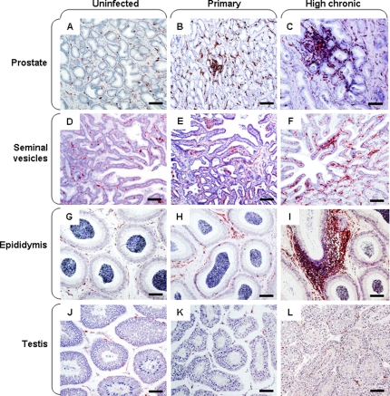
Figure 6. Immune activation in the male genital organs.
Immunohistochemical detection of HLA-DR+ cells in the prostate (A–C), seminal vesicle (D–F), epididymis (G–I) and testis (J–L) of uninfected (A, D, G, J), primary-infected (B, E, H, K) or high chronic macaques (C, F, I, L). Scale bars = 100 µm.

