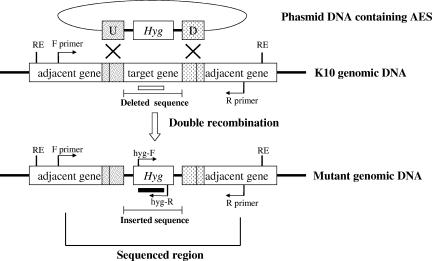
FIG. 1.
Schematic diagram of allelic exchange mutagenesis in M. avium subsp. paratuberculosis. The inserted sequence containing the Hyg gene is the same size in all mutants (1,915 bp), but the size of the deleted sequence varies in the mutants developed in this study (ΔpknG and ΔrelAL, 1,737 bp; ΔrelAS, 873 bp; Δlsr2, 311 bp [Table 2]). Arrows indicate the schematic binding sites and the directions of primers used for PCR identification. The F and R primers are the primers designed to bind outside up- and downstream homologous regions in each mutant. PCRs for ΔrelAS and Δlsr2 were performed with a primer set consisting of F and R primers, because the PCR fragments for the mutant and wild-type strains were clearly distinguished (>1-kb difference). PCRs for ΔpknG and ΔrelAL were performed with specific primer sets consisting of both F and hyg-R primers and hyg-F and R primers because the size differences between the mutant and wild-type strains with the F and R primers were not well distinguished in these cases (178-bp difference). The schematic restriction (RE) and probing (open and filled bars) sites for Southern blot analysis are also shown. Hyg, Hygr gene; U and D, up- and downstream homologous regions; RE, restriction enzyme site; open and filled bars, probes for the deleted gene and the hyg gene, respectively.

