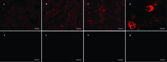
FIG. 5.
Immunocytochemical detection of Ehrlichia in S2 and DH82 cells. Shown are infected S2 cells incubated with E. chaffeensis-specific mouse serum and secondary antibody (A, B, C), infected DH82 cells incubated with E. chaffeensis-specific mouse serum and secondary antibody (D), infected S2 cells incubated with healthy mouse serum and secondary antibody (E), infected S2 cells incubated with secondary antibody only (F), uninfected S2 cells incubated with E. chaffeensis-specific mouse serum and secondary antibody (G), and uninfected DH82 cells incubated with E. chaffeensis-specific mouse serum and secondary antibody (H). Each image was captured at ×20 magnification.

