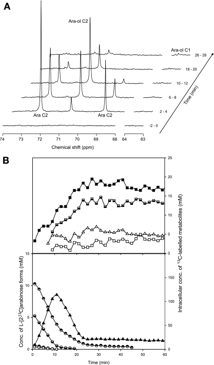
FIG. 1.
l-Arabinose metabolism in suspensions of C. arabinofermentans PYCC 5603T resting cells monitored by in vivo 13C NMR. (A) Sequence of in vivo 13C NMR spectra acquired during the metabolism of l-[2-13C]arabinose (20 mM) at 30°C and under aerobic conditions. Each spectrum represents 2 min of acquisition. Cells were grown in aerobic batch cultures with l-arabinose (20 g liter−1), harvested at exponential growth phase, and suspended in 50 mM phosphate buffer (pH 6.0) to a concentration of around 40 g (dry weight) liter−1. Ara, arabinose; Ara-ol, arabitol. (B) Time course for the consumption of l-[2-13C]arabinose and evolution of the intracellular metabolite pools. Metabolite concentrations were obtained from in vivo 13C NMR data. Symbols: circles, l-arabinose (◓, α-l-[2-13C]arabinopyranoside; ◒, β-l-[2-13C]arabinopyranoside; •, α-l-[2-13C]arabinofuranoside; ○, β-l-[2-13C]arabinopyranoside); triangles, arabitol (▵, C-1 labeled; ▴, C-2 labeled); squares, trehalose (□, C-1 labeled; ▪, C-2 labeled;, C-3 labeled).

