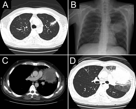
FIG. 1.
(A) CT scan of the chest on admission, showing a small faint patch at the left upper lung field (LULF) and old fibrotic changes at the right upper lung field. (B to D) Follow-up chest radiography 1 month later disclosed a mass lesion at the LULF (B), and a CT scan at the same period (C and D) revealed a mass with a fluid-retaining central cavity, thick irregular wall, and a focal area of consolidation over the LULF.

