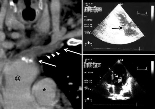
FIG. 2.
(Left) Computed tomographic image generated by curved multiplanar reconstruction showing thrombosis over the left brachiocephalic vein. Arrows, thrombus; arrowheads, peripheral edema of vascular wall; @, superior vena cava; *, aorta. (Right) Transthoracic echocardiographic images. The vegetation sizes were 1.40 cm by 1.30 cm on hospital day 44, when infective endocarditis was first diagnosed (upper right, black arrow), and 2.18 cm by 1.26 cm on hospital day 72, when the blood culture became negative (lower right, white arrow).

