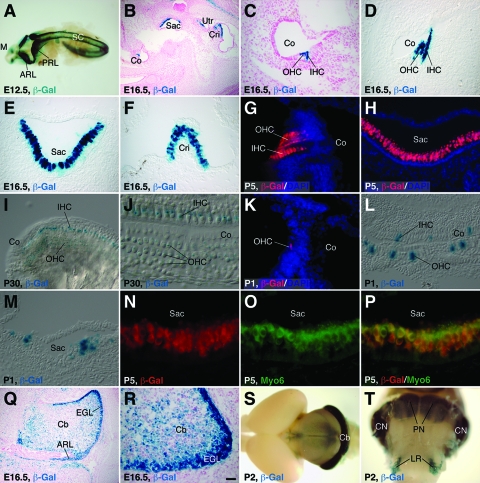
FIG. 2.
β-Gal expression in the inner ear and brain from the Tg1 or Tg3 transgene. (A to J) Using Tg1 animals at the indicated stages, X-Gal staining was performed on whole-mount embryos (A), inner ear sections (B to F), and whole-mount organs of Corti (I and J) or immunofluorescence was carried out on inner ear sections with an anti-β-Gal antibody (G and H). (A) β-Gal activity was seen in the mesencephala, anterior and posterior rhombic lips, and spinal cords at E12.5. (B to J) In the inner ear sections, β-Gal activity was present in hair cells of the cochlear and vestibular systems and this activity was significantly downregulated by P30. (K to M) Inner ear sections of Tg3 neonates were immunolabeled with an anti-β-Gal antibody (K) or stained with X-Gal (L and M). β-Gal activity was observed only in scattered hair cells of the organs of Corti (K and L) and saccules (M). (N to P) Saccular sections from P5 Tg1 animals were labeled by a double-immunofluorescence method with antibodies against β-Gal and Myo6. Note the complete colocalization of β-Gal and Myo6 in hair cells. (Q to T) X-Gal staining of brain sections (Q and R) and whole-mount brains (S and T) from Tg1 animals at the indicated stages. Strong β-Gal activity was seen in the cerebellar granule cells, rhombic lips, PN, CN, and LR nuclei. Sections in panels B, C, Q, and R were counterstained with Fast Red, and those in panels G, H, and K were counterstained with nuclear DAPI. ARL, anterior rhombic lip; Cb, cerebellum; Co, cochlea; Cri, crista; EGL, external granule layer; IHC, inner hair cell; M, mesencephalon; OHC, outer hair cell; PRL, posterior rhombic lip; Sac, saccule; SC, spinal cord; Utr, utricule. Scale bars, 100 μm (B and Q), 25 μm (C to I, K to M, and R), 12.5 μm (J), and 10 μm (N to P).

