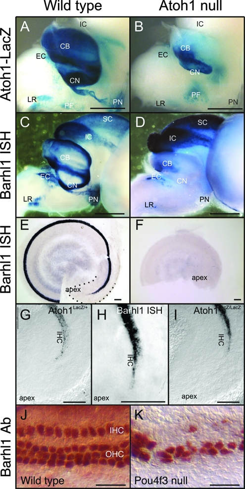
FIG. 6.
Effect of Atoh1 and Pou4f3 inactivation on Barhl1 expression in the ear and brain at E18.5. (A and B) As indicated by the β-Gal activity expressed from the knock-in lacZ reporter, Atoh1 is expressed at high levels in the cerebellum (CB), as well as in the LR nucleus, the PN, the CN, and the EC and perifacial (PF) nuclei (A). In the Atoh1 null brain (B), there is some expression of the lacZ reporter, in particular in the perifacial neurons. However, there is a great loss of neurons and/or lacZ expression in the cerebellum and several nuclei, most notably the PN and CN. IC, inferior colliculus. (C and D) Barhl1 expression in the wild type (C) follows, to some extent, the expression of Atoh1 and is found in the cerebellum and the LR nucleus, PN, and EC nucleus and in some cochlear neurons. Barhl1 is also found in neurons that do not express Atoh1, such as those in the inferior colliculus and the superior colliculus (SC). Some neurons (perifacial nuclei) that are positive for Atoh1 do not show any Barhl1 in situ hybridization (ISH) signals. In the absence of Atoh1 (D), there is a complete loss of Barhl1 expression in the CN and PN and only residual expression in the EC and LR nuclei and the cerebellum. As expected, in areas (interior and superior colliculi) where there is no Atoh1 expression, there is no loss of Barhl1 signals. (E and F) In the ear (E), Barhl1 expression is strong but has not yet reached the cochlear apex at E18.5 (dotted line). Atoh1 null mutant ears (F) show no indication of Barhl1 expression, despite the fact that undifferentiated Atoh1-lacZ-positive cells exist in the apex (I). (G to I) Barhl1 upregulation very closely follows the upregulation of Atoh1, including the absence of expression in the apex and initial upregulation in inner hair cells (IHC) (G and H). In contrast, β-Gal expression in Atoh1 null mice is first detected in the equivalent of outer hair cell (OHC) precursors in the apex (I). (J and K) In the wild-type cochlea, immunostaining with a Barhl1 antibody (Ab) reveals Barhl1 protein expression in the inner and outer hair cells (J). This expression remains strong in residual hair cells of the Pou4f3 null cochlea (K). Scale bars, 1 mm (A to D), 100 μm (E to I), and 25 μm (J and K).

