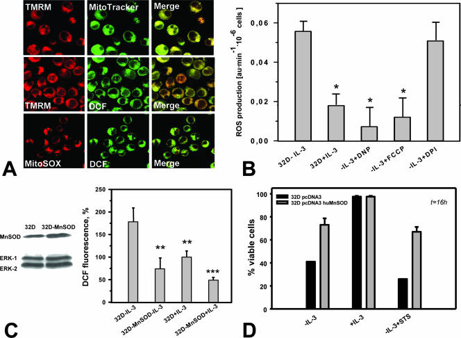
FIG. 3.
ROS production in IL-3-deprived 32D cells occurs primarily at mitochondrial sites. (A) The mitochondrial membrane potential-sensitive dye TMRM fully colocalized with the mitochondrial marker MitoTracker Green (top) but also with DCF-DA fluorescence (green) (middle). Visualization of ROS by the superoxide-specific indicator MitoSOX Red (red) demonstrated colocalization with ROS as detected by DCF-DA (DCF) (bottom). (B) Treatment with either of the mitochondrial uncouplers FCCP (5 nM) and DNP (2 μM) was associated with significantly reduced ROS production (P < 0.05; n = 3), whereas the inhibitor of NADPH oxidase diphenyleneiodonium chloride (DPI) (1 μM) had no effect. au, arbitrary units. (C) Western blot analysis of MnSOD-transfected 32D cells shows elevated expression of MnSOD (left). MnSOD overexpression significantly protects growth factor-deprived 32D cells from excessive ROS production as measured by DCF-DA (DCF) fluorescence (percent control 32D-plus-IL-3 cells [right]). Symbols: **, P < 0.01; ***, P < 0.001 (n = 3). (D) MnSOD overexpression protects 32D cells from cell death induced by IL-3 withdrawal or by STS (7.5 nM).

