FIG. 3.
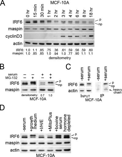
IRF6 protein expression and phosphorylation are regulated by the cell cycle in a proteasome-dependent manner. (A) Western blot analysis depicting changes in IRF6 phosphorylation and expression upon addition of serum to synchronized MCF-10A cells. Time 0 (0 h) represents 48 h of serum starvation, with each subsequent time point indicating time elapsed following serum addition. Densitometry represents the ratio of IRF6 or maspin to actin. (B) Western blot analysis demonstrating the effects of MG-132 on IRF6 expression and phosphorylation in the presence and absence of serum. (C) Coimmunoprecipitation of whole-cell lysate from MCF-10A cells either serum-starved for 48 h (−serum) or stimulated for 1 h with growth medium (+serum). Complexes were immunoprecipitated (IP) with antiubiquitin antibody, and the resulting elution was analyzed by SDS-PAGE and Western blot probed for IRF6. (D) Western blot analysis of IRF6 expression and phosphorylation following various mitogenic stimuli. Growth medium consisted of Dulbecco's modified Eagle's medium-F12 (1:1) supplemented with 5% horse serum, EGF (20 ng/ml), hydrocortisone (0.5 μg/ml), and insulin (10 μg/ml). “I/H/E” represents the addition of insulin, hydrocortisone, and EGF at the same concentrations used in the growth medium but in the absence of serum. The serum supplement MitoPlus was used at 1:1,000. Bovine serum and charcoal-dextran-stripped serum (hormone depleted) were used at 10%. p, phosphorylated; np, nonphosphorylated; +, present; −, absent.

