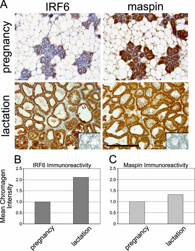
FIG. 4.
IRF6 expression is maximal during lobuloalveolar differentiation (lactation). (A) Immunohistochemistry demonstrating increased IRF6 immunoreactivity during lactation compared to immunoreactivity during pregnancy. Wild-type C57/Black6 mice were harvested at mid-pregnancy and 7 days postparturition. Expression is visualized by the staining intensity of DAB. Slides are counterstained with Mayer's hematoxylin. The negative control inset represents staining with secondary antibody only. Slides, including the negative control, represent serial sections. Bar represents 200 μm. Total magnification is ×200. Pictures were acquired with a Leica DM 4000B microscope mated to a Leica DFC480 charge-coupled-device camera. (B and C) The relative mean intensity of DAB indicating IRF6 or maspin was quantified by using a CMYK color model adapted from a model by Pham et al. (25), based on the chromogen intensity determined from the yellow channel of a CMYK color image.

