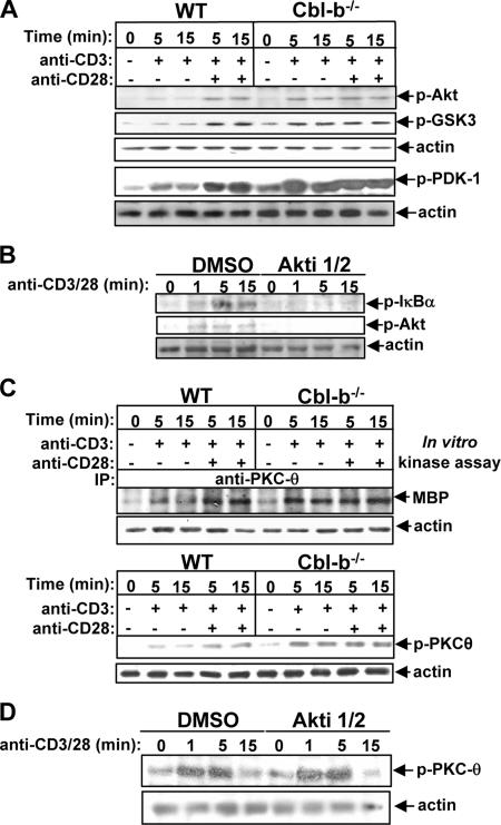
FIG. 2.
Cbl-b down-regulates TCR-induced Akt and PKC-θ activation in primary mouse T cells. (A) WT and Cbl-b−/− T cells were stimulated as in Fig. 1 and lysed. The cell lysates were analyzed with anti-phospho-Akt (Thr308) Ab, anti-phospho-GSK Ab, and anti-phospho-PDK-1 Ab, and reprobed with antiactin Ab. (B) BALB/c T cells were pretreated with Akt inhibitor for 15 min, stimulated with anti-CD3 and anti-CD28 Abs for 1, 5, and 15 min, and then lysed. The cell lysates were analyzed with anti-phospho-IκBα and anti-phospho-Akt Abs. The membrane was reprobed with antiactin Ab. (C) The cell lysates from A were immunoprecipitated with anti-PKC-θ, and the kinase activity associated with PKC-θ immunoprecipitates was determined by in vitro kinase assay using MBP as a substrate, or alternatively, analyzed by anti-phospho-PKC-θ Ab and reprobed with antiactin Ab. (D) Inhibition of Akt does not affect TCR/CD28-induced PKC-θ activation. BALB/c T cells were treated with 10 μM Akti 1/2 for 30 min, stimulated with anti-CD3 and anti-CD28 Abs for 1, 5, and 15 min, and lysed. The phosphorylation of PKC-θ was detected by anti-phospho-PKC-θ Ab, and the membrane was stripped and reprobed with antiactin Ab. Data are from one of four independent experiments. −, absence of; +, presence of.

