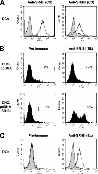
FIG. 1.
SR-BI and SR-BII expression on human DCs. (A) SR-BI and SR-BII expression on human monocyte-derived DCs. Following fixation and permeabilization, DCs were incubated with rabbit anti-SR-BI (NB 400-104) and anti-SR-BII (NB 400-102) polyclonal antibodies directed against the SR-B cytoplasmic domain and subsequently stained with allophycocyanin-conjugated goat anti-rabbit IgG. Cells stained with the secondary antibody alone served as negative controls (gray-shaded curves). The x and y axes show mean fluorescence intensities and relative numbers of stained cells, respectively. (B) Specific binding of mouse anti-human SR-BI to SR-BI expressed in CHO cells. Anti-SR-BI polyclonal serum directed against the SR-BI extracellular loop was raised by genetic immunization of BALB/c mice with a plasmid carrying the full-length human SR-BI cDNA. CHO cells were transfected with pcDNA-SR-BI or control vector (pcDNA). Flow cytometry of SR-BI-transfected CHO cells incubated with mouse anti-human SR-BI polyclonal serum and PE-conjugated anti-mouse IgG demonstrated specific interaction of anti-SR-BI antibodies with human SR-BI. Numbers inside the panels represent the percentage of positively stained cells in relationship to the total number of cells. (C) Detection of cell surface SR-BI on DCs by anti-SR-BI. DCs were incubated with anti-SR-BI or preimmune serum and subsequently stained with PE-conjugated anti-mouse IgG. Cells stained with the secondary antibody alone served as negative controls (gray-shaded curves). CD, SR-BI/II cytoplasmic domain; EL, SR-BI extracellular loop; FL4/2-H, fluorescence 4/2-height.

