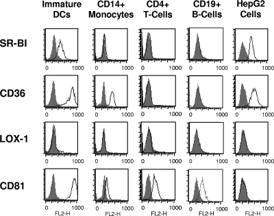
FIG. 2.
SR expression on DCs and other cell types. Cell surface expression of SR was determined by flow cytometry using antibodies directed against SR-BI, CD36, LOX-1, or control antibody and preimmune serum. In addition, cells were stained for CD81 expression using a monoclonal anti-human CD81 antibody. Histograms corresponding to cell surface expression of the respective cell surface molecules (open curves) are overlaid with histograms of cells incubated with the appropriate isotype control (gray-shaded curves [NC]). FL2-H, fluorescence 2-height.

