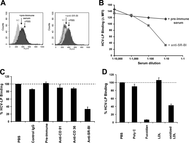
FIG. 5.
HCV-LP binding to human DCs is mediated by SR-BI. (A) DCs were preincubated with anti-SR-BI, preimmune serum, or PBS, and HCV-LP binding to DCs was determined by flow cytometry using a monoclonal anti-HCV E2 antibody and PE-conjugated anti-human IgG. The negative control (NC) histograms represent the results for DCs incubated with an insect cell control preparation. The x and y axes show mean fluorescence intensities and relative numbers of stained cells, respectively. (B) Concentration-dependent inhibition of HCV-LP binding to DCs by anti-SR-BI. Values are shown as net mean fluorescence intensity (Δ MFI) of duplicate measurements. (C) Specific inhibition of cellular HCV-LP binding by anti-SR-BI. Prior to the addition of HCV-LPs, DCs were preincubated with anti-CD36, anti-SR-BI, anti-CD81, control IgG, or preimmune serum. Cellular HCV-LP binding was determined as described above. Data are shown as percent HCV-LP binding (means ± standard deviations of the results from three experiments) relative to HCV-LP binding in the absence of antibodies (100%). (D) Inhibition of cellular HCV-LP binding by SR-B ligands. HCV-LP binding to DCs was determined in the presence of SR ligands fucoidan (1 μg/ml) and oxidized LDL (10 μg/ml) or the control ligands poly(C) (1 μg/ml) and LDL (10 μg/ml). Data are shown as percent HCV-LP binding (means ± standard deviations of the results from three independent experiments) relative to HCV-LP binding in the absence of ligands (100%). FL2-H, fluorescence 2-height.

