FIG. 1.
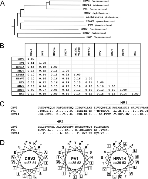
Genetic analysis of picornavirus 2B proteins. (A) Phylogenetic tree of the 2B proteins constructed with Clustal W software. The Blosum62 similarity matrix was used to perform sequence alignment analysis. The phylogenetic tree shows that the 2B proteins of CBV3, PV1, and HRV14 are grouped together, whereas the 2B proteins of viruses from other genera are more distantly related. (B) Identity matrix for 2B proteins. Identity was calculated using the Blosum62 similarity matrix. (C) Pairwise alignment of CBV3, PV1, and HRV14 2B proteins. All three proteins contain two hydrophobic regions (HR1 and HR2) spaced by a 5-aa hydrophilic sequence. Dots represent residues that are identical to those in CBV3 2B. Dashes indicate gaps in the alignment. (D) Top view of the amphipathic α-helices of the first hydrophobic region of CBV3, PV1, and HRV14 2B proteins. Note that all three proteins contain a hydrophobic backbone and a hydrophilic face that contains three cationic residues. Hydrophobic residues are boxed. CBV3, coxsackievirus B3; HRV14, human rhinovirus 14; PV1, poliovirus 1; FMDV, foot-and-mouth disease virus; EMCV, encephalomyocarditis virus; HpaV2, human parechovirus 2; PTV, porcine teschovirus; ERBV, equine rhinitis B virus; HAV, hepatitis A virus.

