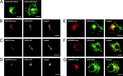
FIG. 4.
Analysis of protein trafficking in cells expressing picornavirus 2B proteins. BGM cells were transfected with the VSV-G-GFP construct (A) or cotransfected with VSV-G-GFP and the picornavirus 2B-myc constructs (B to G). Cells were incubated at 40°C for 18 h to accumulate VSV-G-GFP in the ER and subsequently incubated at 32°C for 2 h (B to G). Cells were fixed, stained with the anti-c-myc antiserum, and processed for CLSM analysis. (A) Accumulation of VSV-G-GFP in the ER upon incubation at 40°C (left) and on the plasma membrane upon additional incubation at 32°C (right). VSV-G-GFP accumulated in the Golgi complex upon incubation with CBV3 2B (B), PV1 2B (C), and HRV14 2B (D). VSV-G-GFP was exposed on the plasma membrane in cells expressing HAV 2B (E), FMDV 2B (F), or EMCV 2B (G). Bars = 10 μm.

