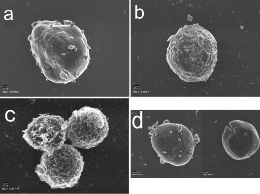
FIG. 7.
Phases of cyst wall formation as detected by scanning electron microscopy. (a) The rounded trophozoite with cellulose patches on the cell surface appears approximately 12 h after the induction of encystation. (b) Immature precyst with a continuous cell wall (approximately 24 h after the induction of encystation). A single layer of the cell wall is indicated by arrows. (c) Three mature cysts. The wrinkled exocyst is clearly recognizable. (d) RNAi-treated acanthamoebae were not able to form a mature cyst. Mag, magnification; 12.00 K X, ×12,000.

