FIG. 6.
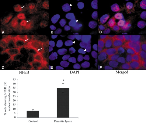
Representative micrographs showing nuclear translocation of NF-κB in intestinal epithelial T84 cells following exposure to B. ratti WR1. T84 cell monolayers were grown on collagen-coated glass coverslips and coincubated with WR1 lysate for 6 h. The monolayers were fixed and incubated first with rabbit anti-NF-κB p50 antibody and then with Cy3-conjugated sheep anti-rabbit secondary antibody. The monolayers were mounted using mounting medium containing DAPI to counterstain the nuclei and visualized by fluorescence microscopy. (A to C) T84 cells exposed to WR1 lysate showed nuclear translocation of the p50 subunit. (D to F) Control cells exhibited normal perinuclear localization of p50. All of the micrographs were obtained at a magnification of ×400. Arrows, p50 localization; arrowheads, nuclei. As shown in the histogram, a significant increase in the percentage of cells showing nuclear translocation of NF-κB p50 could be seen in cells exposed to WR1 lysate. The percentage of cells with NF-κB p50 nuclear translocation was calculated from observations of 200 cells in three independent experiments. The values are means ± standard deviations. *, P < 0.05 versus the control.

