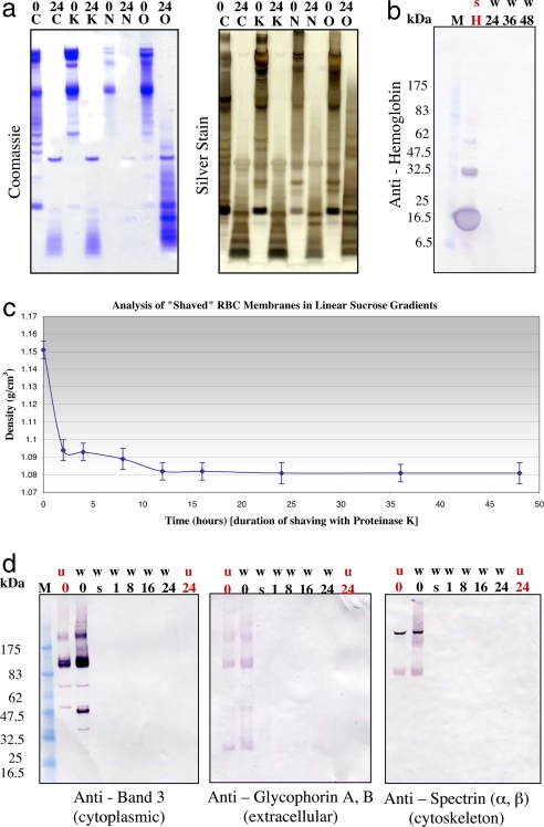
Fig. 1.
Time progression of membrane shaving. (a) Membrane washes. RBC open ghost membranes were washed in 1 M KCl (K), 10 mM NaOH (N), or 100 mM Na2CO3 (O), and then shaved with 3.0 mg/ml proteinase K in the respective wash buffer for 24 h (C, control, membranes were not washed). The membrane samples were then run on 1D SDS/PAGE gels and subjected to standard Coomassie and silver staining procedures. 0, no shaving with proteinase K; 24, membranes were shaved with 3.0 mg/ml proteinase K for 24 h. (b) Elimination of contamination from hemoglobin. RBC open ghost membranes washed (w) in 10 mM NaOH were shaved with 3.0 mg/ml proteinase K in 10 mM NaOH for various lengths of time up to 48 h. The membrane samples were then run on 1D SDS/PAGE gels and subjected to standard immunoblot analysis by using anti-hemoglobin (Sigma, H4890). Lane M, markers; lane H, hemoglobin supernatant (positive control for hemoglobin). (c) Time progression of membrane shaving by using buoyant density measurements from equilibrium centrifugation in linear sucrose gradients. RBC plasma membrane open ghosts were progressively shaved by proteolytic digestion with 3.0 mg/ml proteinase K. The proteolytic digestion was stopped with 5 mM PMSF as a function of time. At each time point, buoyant density measurements were made by using equilibrium centrifugation in continuous, linear sucrose gradients. Membranes were loaded on 5–45% continuous, linear sucrose gradients made by using the BioComp Gradient Master and spun at 215,578 × g for 24 h. Gradients were then fractionated by using the BioComp Gradient Fractionator. The density of sucrose at which the sample band of membranes was found by refractometry (equal to a buoyant density measurement for the membrane itself) was measured by using a temperature-controlled, small-volume, automatic refractometer (Rudolph Research Analytical, J57). The density at each time point was measured independently a total of ten times. (d) Time progression of membrane shaving by using immunoblot analysis. RBC open ghost membranes washed (w) in 10 mM NaOH and unwashed (u) RBC open ghost membranes were shaved with 3.0 mg/ml proteinase K in 10 mM NaOH for various lengths of time up to 24 h. 0, control, no proteinase K; s, shaved for 5 s. The membrane samples were then run on 1D SDS/PAGE gels and subjected to standard immunoblot analysis. Three different antibodies were analyzed in the immunoblot analysis including anti-band 3 (Sigma B9277) (cytoplasmic), anti-glycophorins A and B (Sigma G7650) (extracellular), and anti-spectrin (α and β) (Sigma S3396) (cytoskeleton). Lane M, markers.

