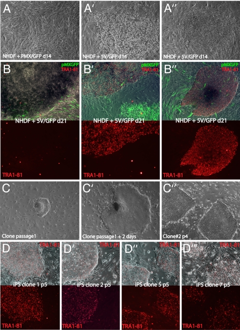
Fig. 1.
iPS clones share HESC morphology. (A′) Heterogeneous morphology of colonies of NHDF1 infected with empty pMX virus and GFP-containing pMX viruses in a 5:1 ratio (pMX/GFP) or a combination of six pMX viruses each carrying one of the five defined transcription factors or GFP (5V/GFP), at day 14 after infection in phase contrast. (B–B″) Phase-contrast images of different colonies from 5V/GFP transduced cultures merged with live TRA-1–81 staining (red) and GFP fluorescence derived from the pMX-GFP virus (green) (Upper) and the TRA-1–81 channel separately (Lower) from cultures transduced with 5V/GFP. Note that only a minor proportion of colonies are TRA-1–81-positive as seen in B and B′. The staining in TRA-1–81-positive colonies was indistinguishable from that obtained with HESC (data not shown). (C–C″) Phase-contrast images of iPS clones at different passages. (D–D‴) “Live” Tra-1–81 staining and merge with phase-contrast appearance of indicated iPS clones at passage 5.

