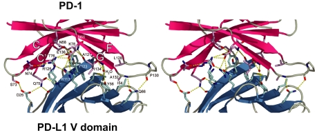
Fig. 2.
The PD-1/PD-L1 interface. Shown is a stereoview of the PD-1/PD-L1 interface showing side chains of residues on β-strands (CC′FG) of PD-1 (red) and on β-strands (GFCC′, left to right) of PD-L1 (blue) that make contacts. Interacting PD-1 side chains (pink) and PD-L1 (light blue) are shown; for clarity a few side chains are not shown. Dotted lines (yellow) indicate hydrogen bonds formed in the interface and with a water molecule.

