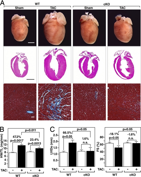
Fig. 2.
Diminished hypertrophy of PKD1 cKO mice after TAC. (A) Hearts from WT and PKD1 mutant mice subjected to either a sham operation (WT and PKD1 cKO, n = 6) or TAC (Top; WT, n = 12; PKD1 cKO, n = 11). Histological sections stained with H&E (Middle) or Masson's trichrome to detect fibrosis (Bottom). (Scale bars: Top and Middle, 2 mm; Bottom, 40 μm.) (B) Heart weight/tibia length (HW/TL) ratios (±SEM) of WT and PKD1 cKO mice were determined 21 days after TAC. (C) PKD1 cKO mice display less left ventricular dilation during systole (LVIDs) and a less pronounced decrease in fractional shortening (FS) in response to TAC than WT mice.

