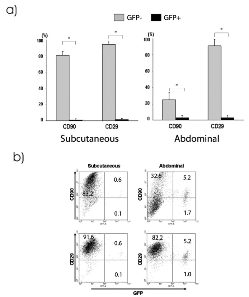
Figure 6.
Surface markers expression of cultured, passage three ASCs from GFP+ radiation chimera. a) ASCs obtained from fat tissues of radiation chimera were analyzed by flow cytometry. CD29 and/or CD90 expressing ASCs were mostly GFP negative; however, small numbers of GFP positive bone marrow-derived ASC were detected. Of note, ASCs from the abdominal fat and subcutaneous fat differ in the expression of CD90. Data is mean ± SD of 3 experiments, * = p ≤ 0.05. b) Representative scattergrams showing GFP+ CD29+/CD90+ ASCs. The intensity of the expression of CD29 and CD90 on GFP+ ASCs is lower than that expressed on GFP− ASCs. The abdominal and subcutaneous fats were obtained from radiation chimera at 161 days after bone marrow transplantation.

