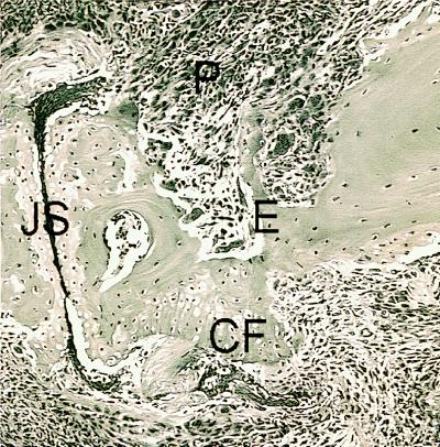
Figure 3.
Histopathology of an arthritic joint from a DR4 and human CD4 transgenic/I-Abβ0/0 mouse immunized with CII. Hematoxylin-eosin staining of a distal interphalangeal joint obtained 60 days after CII immunization. Both inflammatory pannus tissue (P) eroding the bone (E) and cartilage surface and new cartilage formation (CF) are seen. The joint space (JS) is filled with fibrin.

