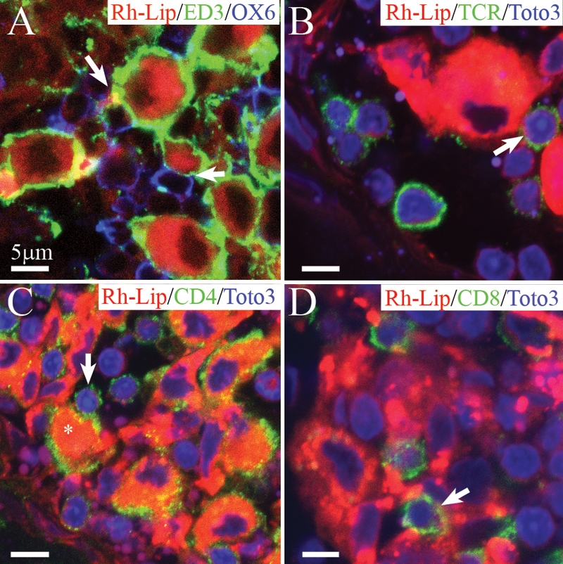
Figure 4.

Biodistribution of rhodamine-conjugated-liposomes in cervical LN in healthy rats 24 h following IVT injection of VIP-Rh-Lip. A: Following IVT injection, VIP-Rh-Lip (red) are located within subcapsular sinus macrophages stained with ED3 (green). OX6-positive cells in blue are located in close proximity (arrow) with ED3-positive macrophages containing VIP-Rh-Lip. B: T lymphocytes recognized by their TCR expression (green) are localized in contact (arrow) with subcapsular macrophages containing VIP-Rh-Lip (red). C: Subcapsular sinus macrophages (asterisk) containing VIP-Rh-Lip (red) express CD4 in green and are in contact with CD4-positive lymphocytes (arrow). D: CD8-positive T lymphocytes (green) are also adjacent with VIP-Rh-Lip (red)-bearing macrophages. In B, C, and D nuclei are stained in blue with Toto3®-iodide. All bars represent 5 μm. Confocal microscopy optical sections are 1.5 μm in all images. Representative images of four experiments performed on cervical LN from four rats.
