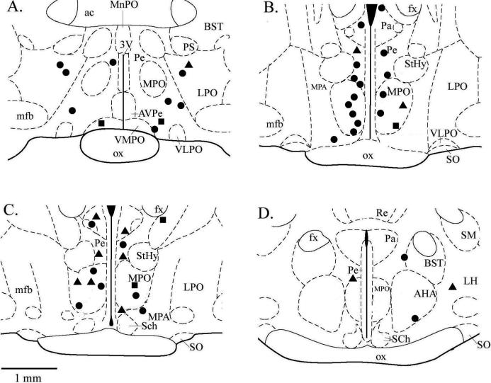
Fig 6. The electrode locations for recordings of single neuron activity in response to temperature and Cirazoline.
Section diagrams are shown in the coronal plane and ordered from rostral to caudal, beginning with the upper left section and moving clockwise. Distance from bregma: A = −0.3 mm; B = −0.8 mm; C = −0.92 mm; D = −1.4 mm. Sections were adapted from an atlas of the rat brain (Paxinos and Watson, 1998). Circles = insensitive neurons which showed a significant increase in firing rate, Squares = insensitive neurons that did not show a change in firing rate, Triangles = warm sensitive neurons which showed a significant decrease in firing rate. 3V, third ventricle; ac, anterior commissure; AHA, anterior hypothalamic area; AVPe, anteroventral periventricular nucleus; BST, bed nucleus stria terminalis; fx, fornix; LH, lateral hypothalamus; LPO, lateral preoptic area; MnPO, median preoptic nucleus; MPO, medial preoptic nucleus; MPA, medial preoptic area; mfb, median forebrain bundle; ox, optic chiasm; Pa, paraventricular nucleus; Pe periventricular nucleus; PS, parastrial nucleus; Re, reunions thalamic nucleus; Sch, suprachiasmatic nuceus; SM, stria medullaris of thalamus; SO, supraoptic nucleus; StHy, striohypothalamic nuc.; VLPO, ventrolateral preoptic area; VMPO, ventromedial preoptic area.

