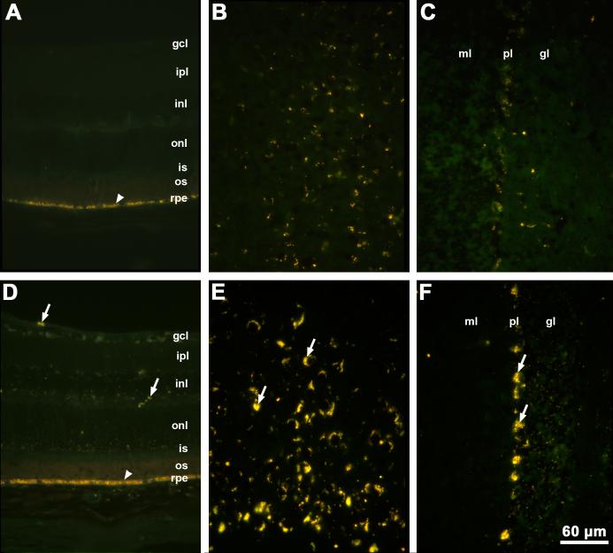
Fig. 2.
Fluorescence micrographs of cryostat sections of retina (A), cerebral cortex (B) and cerebellum (C) from a 12 month old normal C57BL/6J mouse and of retina (D), cerebral cortex (E), and cerebellum (F) from a 12 month old Cln3-/- mouse. Arrows indicate autofluorescent storage material. Arrowheads indicate retinal pigment epithelium with normal autofluorescent age pigment. Bar in (F) indicates magnification in all six micrographs. Layers of the retina are indicated by gcl: ganglion cell layer; ipl: inner plexiform layer; inl: inner nuclear layer; onl: outer nuclear layer; is: photoreceptor inner segments; os: photoreceptor outer segments; rpe: retinal pigment epithelium. Layers of the cerebellum are indicated by ml: molecular layer; pl: Purkinje cell layer; gl: granular layer.

