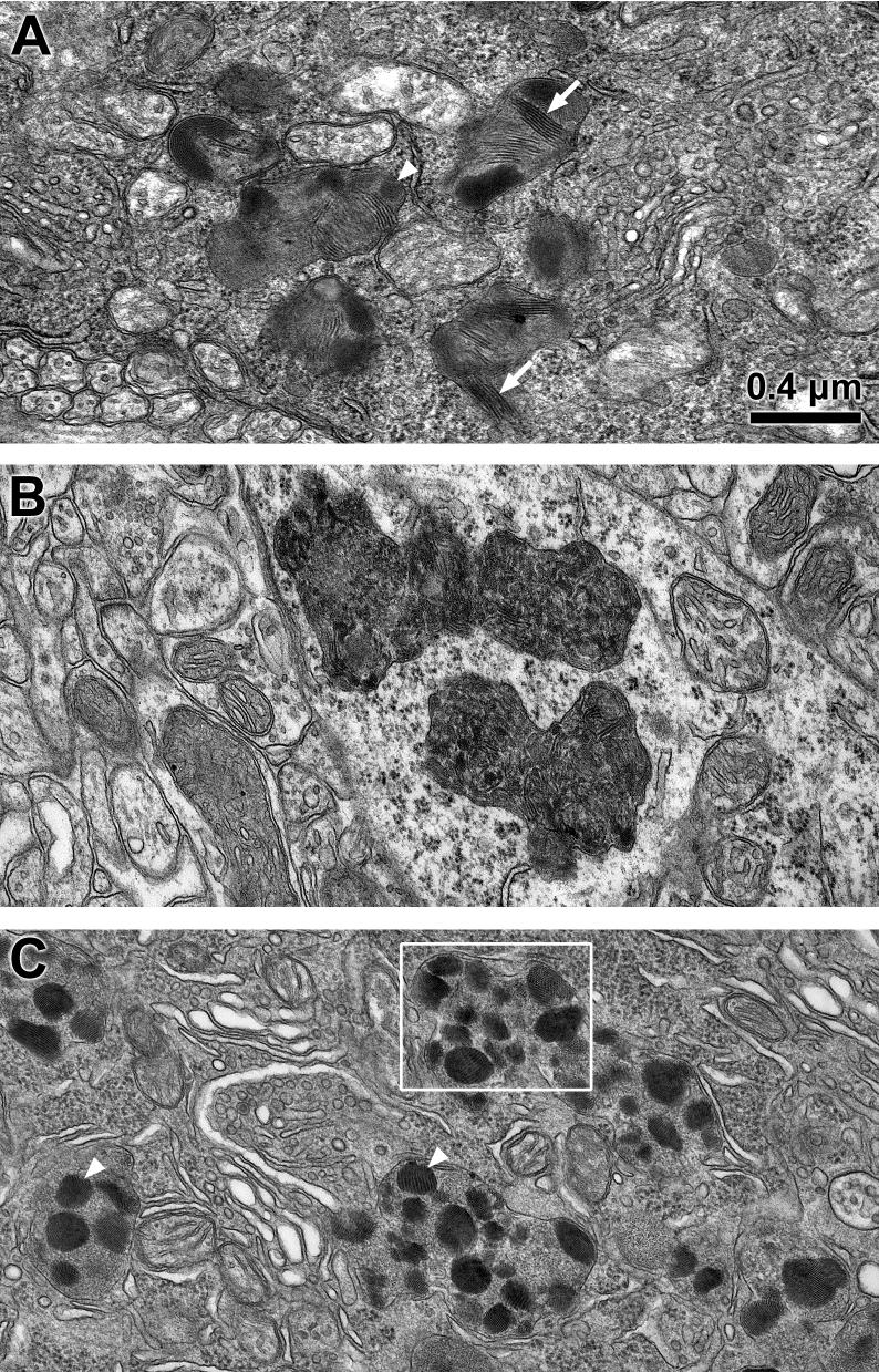
Fig. 4.
Electron micrographs of storage bodies (s) in a retinal ganglion cell (A), cerebral cortex neuron (B) and cerebellar Purkinje cell (C) of a 24 month old Cln3-/- mouse. Storage bodies contained membrane-like (arrows) and crystalline (arrowheads) inclusions. Box in (C) indicates area shown at higher magnification in Fig. 5. Bar in (A) indicates magnification of all three micrographs.

