
FIG. 2.
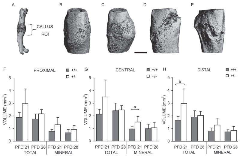
Fracture callus μCT analysis. (A) μCT image of fractured femur with approximate ROI used for volumetric analysis labeled. Representative images of calluses from (B) PFD 21 HIF-1α+/+ mouse; (C) PFD 21 HIF-1α+/− mouse; (D) PFD 28 HIF-1α+/+ mouse; and (E) PFD 28 HIF-1α+/− mouse. Scale bar represents 1 mm. Average total (including mineralized and unmineralized tissue) and mineral volume (only newly formed bone; mm3) were quantified, and bar graphs showing these averages ± SD for (F) proximal; (G) central; and (H) distal regions are presented. ap < 0.03 and bp < 0.04, significant difference between genotypes (Mann Whitney).