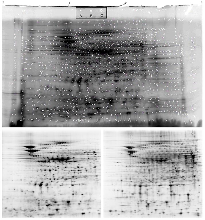
Figure 8. Impact of BODIPY FL-Mal Labeling on Protein 2DGE Mobility.
UPPER: E.coli extracts were split into two fractions: one was labeled with BODIPY FL, and the other alkylated with iodoacetamide. Both were run on separate 2D gels as described in the text. The BODIPY-labeled proteins were imaged on a ProExpress 2D imager, and the unlabeled protein gel stained with Sypro Ruby and likewise imaged. The unmodified tif images were matched using Nonlinear Dynamics Progenesis with no operator modification. The match vectors (white segments) were obtained from the software and the image containing the match vectors overlaid on the BODIPY-labeled protein gel. Inset: A. Circle with average vector diameter (6.6 ± 4.1 pixels in original image); B. 90% confidence interval (10.7 pixels; 1 SD); C. 95% confidence interval (14.8 pixels; 2 SD). LOWER: Unprocessed images of gels used in UPPER. (Left) BODIPY FL-Mal labeled proteins; (Right) Sypro Ruby stained gel. Contrast, brightness, and pixel density of all gel images were adjusted with Photoshop only for publication, after image analysis.

