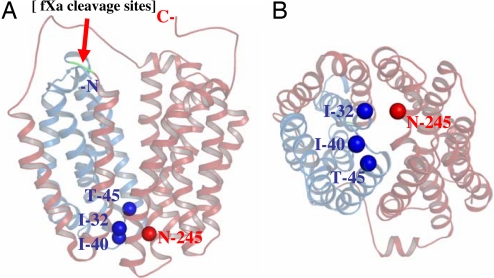
Fig. 1.
X-ray crystal structure of LacY (PDB ID 2V8N). Cα of Ile-32 (helix I), Ile-40 (loop I/II), and Thr-45 (helix II) are presented as dark blue spheres, and the Cα of residue N245 (helix VII) is presented as a red sphere. The N-terminal four helices (N4) and the C-terminal helices (C8) are shown in blue and red, respectively, and separated by tandem factor Xa protease sites (Left; green), as indicated by the red arrow. (A) Side view. (B) Periplasmic view.

