Fig. 4.
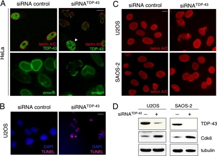
Loss of TDP-43 results in nuclear membrane deformation and apoptosis. Immunofluorescence microscopy of siRNATDP-43 compared with control-treated cells is shown. (Scale bar, 10 μm.) (A) HeLa cells were stained with antibodies against lamin A/C and TDP-43 (red and green, Upper). Arrowhead points to a cell that escaped siRNA silencing. (Lower) Emerin-labeled nuclear membrane. Similar nuclear shape deformation was observed upon detection of lamin B and Lap2β. (B) U2OS cells were assayed for DAPI and TUNEL staining to detect apoptosis. (Scale bar, 10 μm.) (C) The nuclei of U2OS and Saos-2 cells were visualized by lamin A/C detection after TDP-43 depletion. (D) Down-regulation of TDP-43 in U2OS and Saos-2 cells and the consequent Cdk6 increase were verified by immunoblotting. Tubulin was used as loading control.

