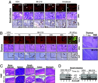
Fig. 5.
MI-219 does not induce tissue damage in normal mouse tissues. (A) Apoptosis in normal small-intestine crypts from mice (three mice per group), treated with a single dose of MI-219 or irinotecan, was examined by TUNEL and H&E staining. p53 accumulation was analyzed by IHC. (B) BALB/c mice (three mice per group) were treated with MI-219 or IR. TUNEL, H&E, and p53 IHC staining were performed in thymus. In addition, H&E staining of thymus was also performed after 7 days of treatment with MI-219. (C) Six nude mice were treated with MI-219 (300 mg/kg BID) for 14 days, and H&E staining of normal mouse tissues was performed. (D) Western blot analysis of p53 activation in small intestine.

