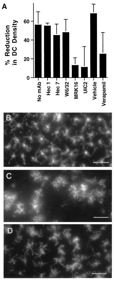
Figure 5.
Retention of DC in the epidermis after treatment with MDR-1 antagonists. (A) Epidermal sheets were stained with anti-MHC II mAb to enumerate DC before the onset of culture or after 3 days of culture in the absence of mAb (no mAb, n = 7), or in the presence of anti-cadherin 5 mAb hec 1 (n = 3); anti-MHC I mAb W6/32 (n = 3); anti-CD31 mAb hec 7 (n = 3); anti-MDR-1 mAb MRK16 (n = 6); anti-MDR-1 mAb UIC2 (n = 1); verapamil (n = 2); or the vehicle control for verapamil 0.03% methanol (Vehicle, n = 1). n = number of experiments in which each condition was examined. DC were counted from en face examinations of epidermal sheets in 16–20 high-power fields per experiment. Percent reduction in DC density was calculated by comparing the number of DC in cultured explants to the mean number present in a portion of the same skin sample before culture (typically 75 cells/field). (B–D) Photomicrographs show the distribution of DC within the epidermis before culturing of explants (B), after 3 days of culture under control conditions (no mAb) (C), and after 3 days of culture in the presence of MRK16 (D). (Bar is 50 μm.)

