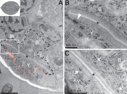
Fig. 2.
Ultrastructural characteristics of Y. (A) Electron micrograph of the rectal area of a newly hatched L1 hermaphrodite showing the B and the Y cells (outlined in black and red, respectively) wrapped around the rectum, both displaying a train-rail-like shape. (Inset) The section of the whole worm from which the rectal area is magnified in A. (B) Blow-up of the boxed area in A, illustrating a C. elegans junction (arrowhead) between the apical membranes of Y and B. (C) Another section of the same L1 animal illustrating a junction between the apical side of the Y cell, or the B cell, and the cuticle of the rectum (arrows). This structure is called a fibrous organelle in C. elegans. An asterisk indicates the rectal slit.

