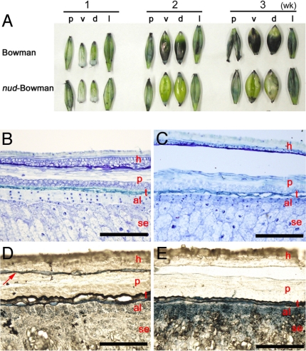
Fig. 4.
Caryopses stained with a lipophilic dye Sudan black B. (A) Hull and dehulled caryopses 1–3 weeks after anthesis. p, palea; v, ventral side of caryopsis; d, dorsal side of caryopsis; l, lemma. Toluidine blue O (B and C) and Sudan black B (D and E) staining of the dorsal-side caryopsis section. (B and D) Bowman. (C and E) nud-Bowman. Red letters are the same as in Fig. 3, except that h indicates lemma. The arrow indicates the lipid layer found only in Bowman. (Scale bars: 200 μm.)

