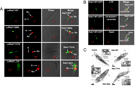
Fig. 2.
Overexpression and localization of LdRab7 in Leishmania. (A) LdRab7 and its mutants were overexpressed in Leishmania promastigotes as described in Materials and Methods. Confocal micrographs showing the localization of LdRab7 and its mutants as GFP fusion proteins. Nucleus (nu) and kinetoplast (k) were stained with propidium iodide. (B) To characterize Rab7 positive compartment in Leishmania, lysosomes (Top) and Golgi complex (Middle) of the LdRab7-GFP overexpressed Leishmania were labeled by incubating the cells with lysotracker red (1 μM) or BODIPY-TR ceramide (5 μM), respectively, at 23°C for 30 min in serum-free M199 medium. Similarly, early endosomal compartment was visualized by anti-Rab5 antibody subsequently probed with goat anti-mouse Alexa Fluor 488-labeled second antibody after fixing the cells with formaldehyde (Lower). Yellow indicates the colocalization of Ld Rab7 with indicated compartment in one plane after after Z-stack analysis by confocal microscopy. (C) Morphology of the endocytic compartments in LdRab7 and its various mutants overexpressed Leishmania were determined by electron microscopic analysis as described in Materials and Methods.

