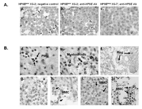
Figure 6. HPSE is expressed by BM environment cells and by a minor subpopulation of myeloma cells.
A. XG-2 and XG-7 cells were stained with control rabbit polyclonal antibodies (Ab) (panel a) or with a polyclonal anti-HPSE Ab at 4.5 μg/ml (panels b,c). Note the dot-like staining signal. B. BM biopsies from patients with MM were stained with the polyclonal anti-HPSE Ab (4.5 μg/ml). Panels d–f: environment cells in the BM of one patient representative of twenty. PMN, polymorphonuclear cells; Meg, megakaryocytes. Panels g–j: heterogeneous HPSE expression among MMC in the bone marrow of four representative patients (g=patient 1; h=patient 4; i=patient 11; j=patient 20; see Table 3). The brown reaction product indicates the location of polyclonal Ab against HPSE. Original magnification of 1000× (or200× in f).

