Fig. 2.
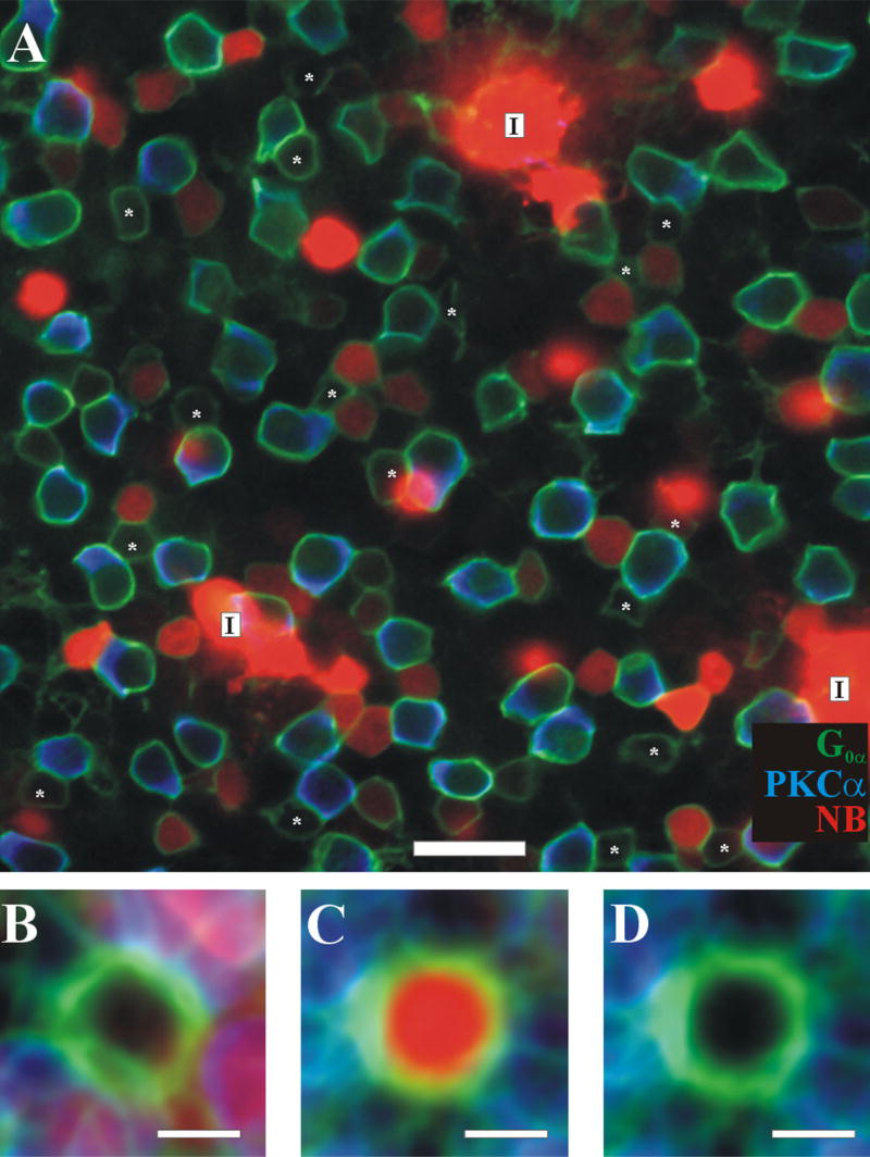
Confocal image of a triple-labeled retina that has a ring of three injected AII amacrine cells. A: Triple-labeled tissue with G0α (green), PKCα (blue), and Neurobiotin (red) filled cells. The letter I represents an injection site. Even though there are three injection sites, there are still cells that are labeled with G0α and have no Neurobiotin in them, which are identified as noncoupled ON cone bipolars (asterisks). There are noncoupled ON cone bipolar cells in the middle of the three injection sites, where Neurobiotin diffusion from the three AIIs is combined, suggesting that poor diffusion does not account for the lack of Neurobiotin labeling in the noncoupled ON cone bipolars. B–D: Same as in Figure 1. Scale bar = 20 μm in A; 5 μm in B–D.

