Figure 1.
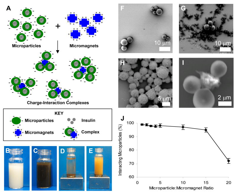
(A) Scheme. Complexes formed through the interaction of negatively charged protein-containing polymer microparticles and positively charged micromagnets. (B)–(E) Particles in suspension: (B) PLGA microparticles alone; (C) Microparticle-micromagnet complexes without external magnet; (D) Complexes with external magnet applied—particles have precipitated; (E) Non-interacting negatively-charged microparticles and negatively-charged micromagnets with external magnet applied—particles remain in suspension. (F)–(I) Scanning electron microscopy (SEM) analysis for the interaction of carboxylate terminated, negatively-charged PLGA microparticles (spherical shape) (~ 4 mm) and positively-charged micromagnets (~ 1 or 6 mm) (irregular shape): (F) PLGA microparticles alone; (G) Non-interacting negatively-charged microparticles and negatively-charged micromagnets; (H) A microparticle-micromagnet complex formed by charge interaction; (I) High magnification of one complex. (J) Mixing of microparticle:micromagnet complexes at various wt:wt ratios, demonstrating that the majority of microparticles were complexed with micromagnets up to a 15:1 mass ratio.

