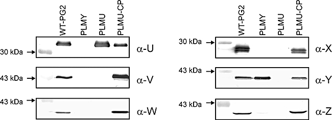
Fig. 4.

Comparative Western blot analysis of whole-cell extracts of M. agalactiae type strain PG2 (WT-PG2), two PLMs (PLMY and PLMU) and xer1-complemented PLMU (PLMU-CP) using six different Vpma-specific pAbs as described in Table 1. Designations of individual pAbs used for each Western blot are indicated in the right margins of each panel whereas relevant protein size standards are shown on the left margins.
