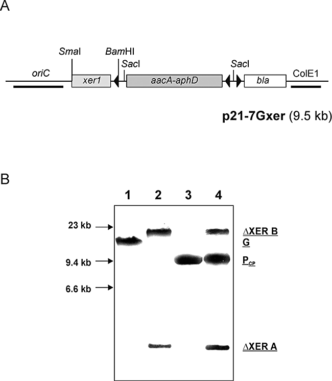
Fig. 6.

Complementation of the wt xer1 gene in PLMU. A. Schematic representation of complementation plasmid p21-7Gxer. Restriction sites used for cloning purposes are as indicated and arrowheads represent direction of transcription; ColE1, E. coli origin of replication; bla, ampicillin resistance gene; aacA-aphD, GentR gene; oriC, M. agalactiae origin of replication. B. Southern blot analysis of xer1-complemented PLMU. ClaI digest of DNA from the complementation clone PLMU-CP (lane 4) is shown in comparison with wt strain PG2 (lane 1), PLMU (lane 2) and complementation plasmid p21-7Gxer (lane 3) after hybridization with a xer1-specific DIG-labelled probe. G represents the 13 kb ClaI fragment of M. agalactiae type strain PG2 whereas ΔXERA and ΔXERB represent the two ClaI fragments from PLMU (as described in Fig. 3); PCP depicts the 9.5 kb linearized complementation plasmid p21-7Gxer. DNA size standards are indicated in the left margin.
