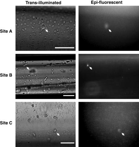
Fig 2.
Paired brightfield and epi-fluorescence images of captured cells immunostained for CD34. Comparison of representative paired brightfield and fluorescent images of adherent cells immunolabelled with an antibody against a hematopoietic stem cell surface marker conjugated to quantum dots revealed blood-borne CD34-bright cells (white arrows) distinctly visible among CD34-dark cells on the capture surface (bar = 50 μm). Quantitative analysis confirmed a sixfold enrichment in the purity of CD34-positive cells on P-selectin surfaces over the proportion found in rat whole blood.

