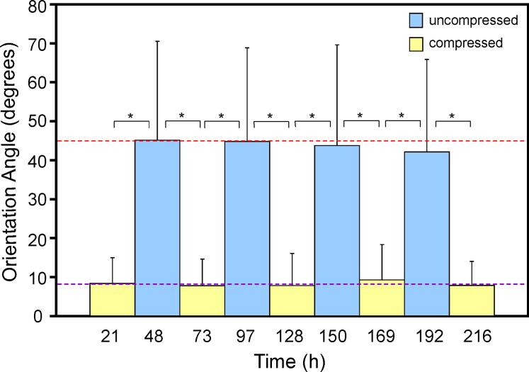
Fig. 1.
Micrographs of the compressed substrate with a comparison image. Larger images are of replicas of a compressed substrate from a top view (top left, optical), angled view (top right, SEM), and cross-sectional view (bottom, SEM). Smaller image is of oxidized, not compressed PDMS for comparison (optical).


