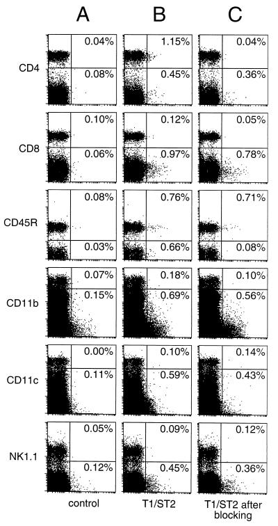
Figure 2.
Expression of T1/ST2 on spleen cells ex vivo. Spleen cells from BALB/c mice were stained with digoxigenized mAb against T1/ST2 (3E10), followed by Cy5-conjugated anti-DIG and PE- or FITC-conjugated mAbs against either CD4, CD8, CD45R, CD11b, or CD11c. B6 spleens were used for the detection of NK1.1+ cells. Gates were set on viable cells according to forward and sideward scatter and exclusion of propidium iodide-binding particles. All samples were incubated with blocking anti-Fcγ-R mAb and purified rat IgG before and during staining with 3E10. Up to 106 cells were acquired to allow depiction of ≈5,000 cells of each leukocyte subset in the upper quadrants of each dot plot. The percentages shown indicate the frequency of T1/ST2+ cells within the different leukocyte subpopulations. (A) Anti-DIG-Cy5 antibody alone. The specificity of the staining for T1/ST2 shown in B was controlled by preincubation of the cells with a 100-fold excess of unconjugated 3E10 as shown in C.

